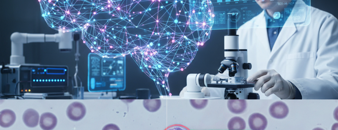
Acute lymphoblastic leukemia (ALL) is one of the most common cancers in children, though it also affects adults. It is a type of blood cancer that develops when immature white blood cells, called lymphoblasts, multiply rapidly and crowd out healthy blood cells. These abnormal cells interfere with normal blood and immune functions, making patients vulnerable to infections, anemia, and other life-threatening complications.
For children, treatments have improved dramatically over the past several decades. Today, the survival rate for childhood ALL is close to 90%. For adults, however, survival rates remain much lower, at only 40–50%. One of the biggest factors influencing these outcomes is how quickly and accurately the disease is detected. Early diagnosis means doctors can begin treatment sooner, improving the chance of survival and reducing complications.
Traditional methods of diagnosing ALL rely on examining blood and bone marrow samples under a microscope. Specialists look for visual signs of abnormal lymphoblasts. While this method is effective, it depends heavily on the expertise of trained professionals, and results can vary depending on the lab, the quality of samples, and even human judgment. This challenge opens the door for new approaches that can make the diagnostic process more consistent, efficient, and widely accessible.
Methods
In this study, researchers developed a computer program that uses artificial intelligence (AI) to identify leukemia in microscope images. The specific AI technique they used is called a Convolutional Neural Network (CNN). CNNs are a type of deep learning model inspired by how the human brain processes visual information. They work by analyzing images in layers, first detecting basic features like edges or shapes, and then combining them into more complex patterns.
To picture how this works, imagine teaching someone to recognize a cat. At first, they notice small features: whiskers, ears, and eyes. Over time, they learn how these features come together to form the bigger picture of a cat. In the same way, the CNN “learns” to recognize what leukemia cells look like by reviewing many microscope images and adjusting its internal system until it can correctly identify the disease.
The CNN used in this study contained five main blocks of layers, with thirteen convolutional (pattern-detecting) layers and five “max-pooling” layers. The pooling layers act like summary steps, condensing the information so the system can focus on the most important features. The model was trained for 30 epochs, meaning it reviewed the entire training dataset 30 times. Each time, it improved its ability to classify images. The researchers also set a batch size of 32, which means the system updated its learning after reviewing 32 images at a time.
Finally, they tested two methods for helping the system learn, called “optimizers.” One of these, known as Adam, turned out to be more effective, helping the CNN adjust its learning process in a smart, efficient way.
Results
Once trained, the CNN was tested on about 1,800 microscope images. The results were striking:
- The system achieved 90% accuracy, meaning it identified the presence or absence of leukemia correctly in 9 out of every 10 cases.
- Its precision was 95%, which means that when it predicted a sample was positive for leukemia, it was correct 95% of the time.
These results suggest that the AI model performs at a level that is both clinically useful and comparable to, or better than, more complex systems developed for similar purposes. Importantly, the CNN achieved this without requiring an overwhelming amount of computing power, making it more practical for real-world settings.
Discussion
Why do these results matter? First, the model could provide much-needed support in places where trained specialists are limited. A clinic in a rural area, for example, may not have a hematologist available to analyze every blood sample. An AI tool like this could serve as an additional layer of screening, ensuring suspicious cases are flagged quickly for further review.
Second, the system provides consistency. Human experts can vary in their interpretations, especially when samples are unclear or borderline. An AI tool can apply the same standards every time, reducing variability in diagnoses.
The researchers also highlight the potential for improvement. While this model used only images, future systems could combine multiple types of patient data—such as genetic information, clinical history, or lab results—to make even more accurate predictions. They also plan to test the model in real hospitals across different regions to ensure it works well outside the laboratory setting.
Conclusion
This study shows how artificial intelligence can play a transformative role in medical diagnostics. Acute lymphoblastic leukemia is a disease where time is critical, as faster and more reliable detection could make a life-or-death difference. By using a Convolutional Neural Network, researchers created a tool that can identify leukemia in microscope images with around 90% accuracy and 95% precision.
Although it is not meant to replace doctors, the system could act as a powerful support tool. In practice, this means quicker diagnoses, earlier treatments, and better outcomes for patients—particularly in places where access to specialists is limited. As AI continues to develop, tools like this may become a standard part of healthcare, bridging gaps in access and bringing high-quality diagnostic support to patients around the world.